NERVES OF UPPER LIMB – MEDIAN, ULNAR, RADIAL NERVES
MEDIAN NERVE:
Root value: C5, C6, C7, C8, T1
It is formed in the axilla by the union of two roots – medial and lateral
Formation in the axilla:
Medial root is form medial cord of brachial plexus ( C8,T1) and lateral root is from lateral cord of brachial plexus ( C5,C6,C7)
The medial root crosses the third part of axillary artery and joins with lateral root to form median nerve which is now lateral to the 3rd part of axillary artery.
v

Course:
In the front of arm
In the cubital fossa
In the front of forearm
In the palm of hand
In the front of arm:
It runs lateral to third part of axillary artery and proximal part of brachial artery.
In the middle of the arm, opposite the insertion of coracobrachialis, the median nerve crosses from lateral to medial side.
It runs along the medial side of lower part of brachial artery and enters the cubital fossa as the medial most content.

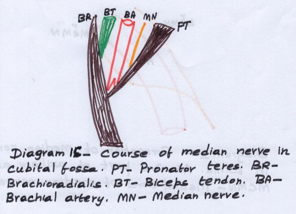
In the cubital fossa:
It is medial to brachial artery in the cubital fossa and leaves the fossa between the two heads of pronator teres.
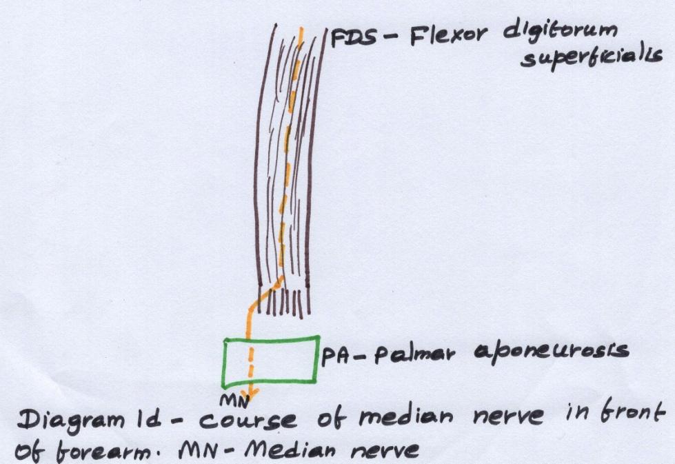
In the front of fore arm:
The median nerve enters the front of forearm under cover of tendinous arch of origin of flexor digitorum superficialis muscle.
It is closely plastered to the under surface of flexor digitorum superficialis muscle.
About 5 cm above the flexor retinaculum, the nerve lies between the flexor carpi radialis and plamaris longus tendon.
It enters the palm of the hand by passing deep to flexor retinaculum of hand.
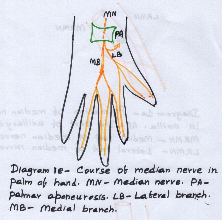
In the palm of hand:
In the palm of hand, the median nerve divides into lateral and medial branches.
The lateral branch gives a recurrent musclular branch to supply the thenar muscles and then divides into three proper digital nerve and the medial branch divides into two common digital nerves
Branches of median nerve:
In the axilla: No branches
In the arm: Nerve to pronator teres just above elbow.
In the fore arm:
Muscular branches- to flexor carpi radialis, palmaris longus, flexor digitorum superficialis
Anterior interosseous nerve- a branch of median nerve supplies flexor pollicis longus, lateral half of flexor digitorum profundus, pronator quadratus
Cutaneous branches: Palmar cutaneous branch just above flexor retinaculum
In the palm:
Muscular branches: to thenar muscles – abductor pollicis brevis, flexor pollicis brevis, opponens pollicis, first and second lumbricals.
Cutaneous branches: Palmar digital nerves to lateral three and half digits
Articular branches: To the elbow, superior and inferior radio ulnar joints and wrist joint.
Vascular branches: To the axillary, brachial arteries

Applied anatomy:
Median nerve is called laborer's nerve as it supplies most of the large flexor muscles of forearm.
Ape like hand: when the median nerve is injured, wasting of thenar muscles and unopposed action of extensor pollicis longus resembles the ape like hand.
Pointing index finger: When the nerve is injured in the middle of forearm, the branch of flexor digitorum superficialis to the index finger is involved, resulting in weakness of flexion of index finger and unopposed extension of that finger – called as pointing index finger.
Carpal tunnel syndrome: Compression of median nerve in the carpal tunnel causes weakness and wasting of thenar muscles and loss of opposition of thumb.
ULNAR NERVE
Root value: C8, T1
Formation: Formed in the axilla as a continuation of medial cord of brachial plexus.
Course:
In the axilla
In the arm – front and back
In the front of forearm
In the palm of hand
In the axilla:
The ulnar nerve is medial to 3rd part of axillary artery deep to the medial cutaneous nerve of forearm.
In the arm:
The ulnar nerve is medial to the upper part of brachial artery
In the middle of arm, it pierces the medial intermuscular septum along with superior ulnar collateral artery and enters the back of am.
In the back of arm, it descends between the septum and medial head of triceps and appears between the medial epicondyle and the olecranon process.
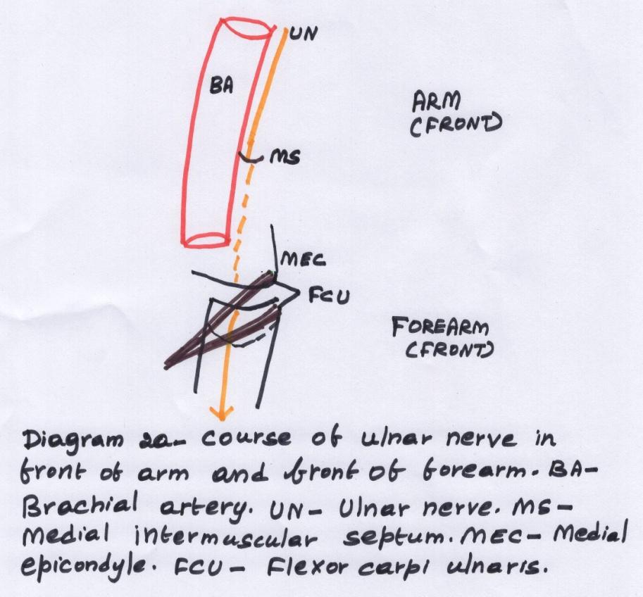
In front of forearm:
The ulnar nerve enters the front of forearm between the two heads of flexor carpi ulnaris
It descends along the medial side of front of forearm accompanied by ulnar artery on its lateral side.
In the upper one third, it is deeply placed and lower 2/3rds, it is superficial on the lateral side of flexor carpi ulnaris.
In the palm of hand:
The ulnar nerve and artery pass superficial to flexor retinaculum of hand.
In the palm of hand, under cover of palmaris brevis, the ulnar nerve divides in to
Superficial terminal branch
Deep terminal branch
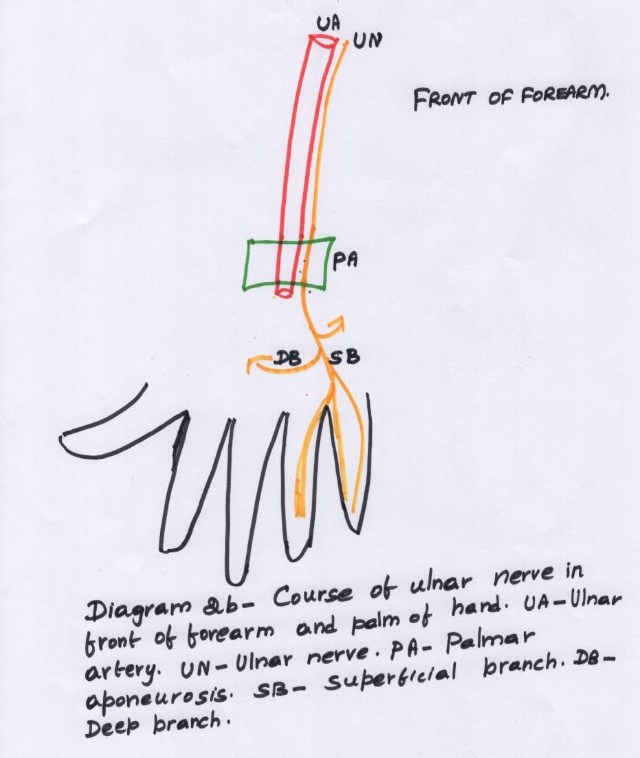
Superficial terminal branch: Supplies the palmaris brevis and divides into digital nerves
Deep terminal branch:
This branch passes deeply between abductor and flexor digiti minimi and pierces opponens digiti minimi
It turns laterally to lodge in a groove below the hook of hamate in the concavity of deep palmar arch.
Branches of ulnar nerve:
In the axilla – no branches
In the fore arm:
Muscular branches: to the flexor carpi ulnaris and medial half of flexor digitorum profundus.
Cutaneous branches:
A palmar cutaneous branch
A dorsal branch to dorsum of hand.
In palm:
Muscular branches: to all intrinsic muscles of hand except three thenar muscles and first and second lumbricals
Cutaneous branches: Superficial terminal branch – gives one proper digital nerve to the medial side of little finger and one common digital nerve to the sides of little and ring fingers.
Articular branches of ulnar nerve: To the elbow, inter carpal, carpometacarpal joints of hand
Vascular branches: To axillary, brachial and ulnar arteries.

Applied anatomy of ulnar nerve:
Cubital tunnel syndrome: compression of ulnar nerve behind the medial epicondyle is called cubital tunnel syndrome.
Manifestations are:
The hand is abducted on flexing the wrist due to unopposed action of flexor carpi radialis.
Medial four fingers cannot be abducted or abducted due to the paralysis of dorsal and palmar interossei.
Claw hand: It is a condition of ulnar nerve injury in which medial four fingers are extended at the metacarpophalangeal joints and flexed at interphalangeal joints.
Wasting of hypothenar muscles and loss of sensation of medial one and half digits and the adjoining medial side of hand in ulnar nerve lesions.
RADIAL NERVE:
It is the continuation of the posterior cord of brachial plexus.
Root value: C5, C6, C7, C8, T1
Course:
In the axilla
In the arm – front and back
In the axilla:
In the axilla, it is posterior to the 3rd part of axillary artery and medial to axillary nerve.
In front of arm:
The radial nerve is posterior to the proximal part of brachial artery.
It leaves the front of arm through lower triangular space accompanied by profunda brachii artery
In back of arm:
The radial nerve along with profunda brachii artery enters the spiral groove and runs downwards and laterally between the lateral and medial heads of triceps.
It pierces the lateral intermuscular septum in the lower part and enters the anterior compartment again.
Front of arm:
It enters the front of arm between brachioradialis and extensor carpi radialis longus laterally and brachialis medially
On reaching the front of lateral epicondyle, the radial nerve divides into superficial and deep terminal branches.

Superficial terminal branch:
This branch is sensory and runs downwards along the lateral side of front of forearm
Accompanied by radial artery on the medial side
About 7cms above the wrist, the nerve winds dorsally around the lateral side of radius and runs in the roof of anatomical snuff box with cephalic vein.
It enters the dorsum of hand superficial to extensor retinaculum
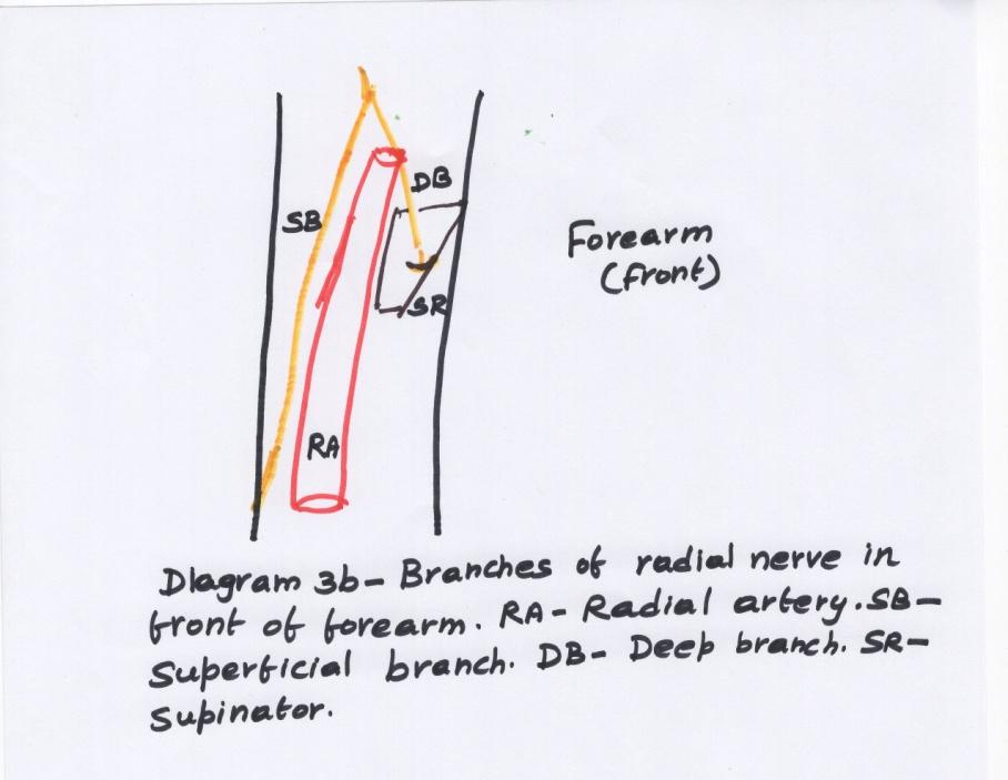
Deep terminal branch
Also known as posterior interosseous nerve.
It pierces the supinator and descends in the back of forearm between the superficial and deep groups of extensor muscles.
It terminates s pseudo-ganglion at the back of wrist.
Branches of radial nerve:
In the axilla:
Muscular branches: to long and medial heads of triceps
Cutaneous: posterior cutaneous nerve of the arm
In the spiral groove:
Muscular branches: To lateral and medial heads of triceps and the branch to medial head supplies the anconeus
Cutaneous: Posterior cutaneous nerve of forearm and lower lateral cutaneous nerve of arm
In the arm:
In the lower part, muscular branches to: brachioradialis, extensor carpi radialis longus and brachialis
Superficial terminal branch:
It is sensory and gives cutaneous nerves five dorsal digital nerves to lateral 2/3 of dorsum of hand and dorsal aspect of lateral three and half digits.

Deep terminal branch:
Muscular branches to the extensor group of muscles of forearm – all the superficial and deep muscles
Applied anatomy:
Saturday night palsy: Injury to radial nerve in spiral groove by placing the outstretched arm on an arm chair under drunken condition is associated with temporary radial nerve palsy.
Wrist drop : When paralyzed, the hand is flaccid and flexed at the wrist
Extension of elbow is last due to the triceps paralysis.


Comments
Post a Comment